Posts

Showing posts with the label Life Science

Eukaryotic Cell

Image
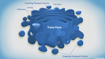
  Eukaryotic Cell Eukaryotic Cell The cells which contain true nuclei are called eukaryotic cells. Eukaryotic cells are found in animals, plants, algae and fungi. They are larger in size as compared to prokaryotic cells. The cells may vary in shape, size and physiology but they all have a typical structure with little variations in number and location of cellular organelles. The eukaryotic cell have a outer covering membrane called plasma membrane / cell membrane. Inside the cell a membrane-bound nucleus is present. Between the nucleus and plasma membrane, cytoplasm is present in which various cellular organelles like mitochondria, golgi complex, centrioles etc can be seen. ✱ Shape: Eukaryotic cells exhibit diversity. They can be spherical, triangular, tubular, cuboidal, polygonal, cylindrical, oval, rounded or elongated. Shape of cells may vary from organ to organ. A single organ may show variations in shape of cell. ✱ Size: Eukaryotic cells are microscopic. They are larger in size

Chromosomes

Image
  (Image Source:  https://pmgbiology.files.wordpress.com/2015/10/chromosomes.jpg?w=300 ) Chromosomes are found inside the nucleus of the cell. The number of chromosomes differ from organism to organism. Humans have 23 pairs i.e. 46 chromosomes. Out of these 23 pairs, 22 pairs are autosomes and 1 pair is of sex chromosomes. Human female have XX as the sex chromosome and human male have XY as the sex chromosome. One set (23 chromosome) is inherited from the maternal (female parent) and another set from the paternal (male parent). Since two sets are present inside the cell, the cell is called as diploid (di=two, ploid=set). All somatic cells of the body are diploid. Gametes (sperm and ovum) contains only one set of chromosomes and hence are called haploid. The number of chromosomes in each somatic cell is the same for all members of a given species.  Chromosome Morphology: (Image Source:  https://microbiologynotes.org/wp-content/uploads/2020/07/types-of-Telomere.jpg ) Chromosomes can be d

Subphylum Urochordata

Image
  (Image Source:  https://scopeblog.stanford.edu/2013/08/04/image-of-the-week-botryllus-organism/ ) The Urochordata also known as Tunicata is a part of Phylum Chordata. General Characteristics of Urochordates: They are marine and sedentary. Their body is enclosed in a test/tunica. Notochord is present in the tail region in larvae (hence urochordate) and disappears in adults. Alimentary canal is complete and are ciliary feeders. Respiration is through numerous gill slits (Stigmata) which opens into the ectoderm lined cavity, the atrium. They show open circulation. They have special corpuscles, vanadocytes (except in Herdmania ) to extract vanadium from sea water and store it in blood vessels. Dorsal tubular nerve chord appears only in larval stage, which in adults is replaced by a single dorsal ganglia. Sense organs are in the form of receptors. Excretion is by nephrocytes, pyloric gland or neural gland. Asexual reproduction is by budding. Mostly hermaphrodite. Their development is indi

Integument

INTEGUMENT: Vertebrate body is covered by a tough, three layered body wall consisting of outer integument, middle muscular layer, and inner coelomic epithelium. It displays important morphological and physiological variations between different vertebrate groups depending on their adaptations for various habitats and mode of life. However, being a protective covering the integument is basically similar in structure in all vertebrates. Histologically skin is made up of two distinct layers. Epidermis: Ectodermal, stratified squamous epithelium, made up of few to several layers of cells. These layers include: Stratum germinativum/stratum malpighi/ stratum cyclindricum: It is the the innermost layer, firmly attached with the dermis and is settled on the basement membrane . It is continuously growing layer which push the new cell layers towards surface. It gets nourishment from dermis. Stratum corneum: It is the outermost partially cornified layer consisting of dead cells. It is made up of p

Reproduction in Amoeba

Image
  Amoeba Amoeba always reproduce asexually. There are different ways of asexual reproduction in Amoeba and they are as follows: 1) Binary Fission: Parent Amoeba divides into two daughter amoebae in favorable conditions. This is known as binary fission. Different phases of mitosis are accompanied with changes in the shape of body. In prophase, body becomes spherical with numerous pseudopodia and nucleus show intranuclear spindle. Chromatids appear as duplicated chromosomes in the nucleus. In metaphase chromosome arrange at the equator. In anaphase daughter chromosomes move towards opposite poles. In telophase. pseudopodia assume normal shape, constriction appear in the middle. One of the daughter retain contractile vacuole and the other acquires new vacuole. Now the parent after binary fission starts living as two individuals which later on grow in size.  Binary Fission in Amoeba 2) Sporulation: Some species of Amoeba reproduce by sporulation. Sporulation begins with breakdown of nuclea

Golgi Apparatus

Image
  Golgi Complex / Golgi Apparatus Golgi apparatus is found in all types of eukaryotic cells except the mammalian RBCs. It is absent in prokaryotes. Golgi apparatus are small sacs which are responsible for transporting, modifying and packaging proteins and lipids. The golgi apparatus was discovered by Camillo Golgi in 1898. He discovered them in the nerve cells. Golgi apparatus has a system of outer flattened cisternae which appears as parallel membranes in the form of an ellipse.  There are two faces known as cis and trans face of golgi apparatus. The cis face lies near the rough endoplasmic reticulum and the trans face lies near to the cell membrane. Substances from the rough endoplasmic reticulum enter through the cis face for processing and they exit from the trans face in the form of smaller vesicles. Functions: Packaging center of cell. Proteins are processed and sorted for their transport. Lipid processing. Secretory proteins like hormones are packed into secretory vesicles for t

Endoplasmic Reticulum

Image
  Endoplasmic Reticulum (ER) Endoplasmic Reticulum (ER) is a system of sac like structures and tubes found in all eukaryotic cells. They are known as sarcoplasmic reticulum in skeletal muscles, Nissl granules in neurons and myeloid bodies in retinal pigment cells. They are absent in RBCs, eggs and embryonic cells. Endoplasmic Reticulum arise by evagination of nuclear membrane. Endoplasmic Reticulum have three parts: Cisternae: It is a sac like, parallel tubules having ribosomes on their surface. It is unbranched. Tubules: It is tube like, irregular, branched and without ribosome. Vesicles: They are round or spherical, without ribosomes Parts of Endoplasmic Reticulum . There are two types of endoplasmic reticulum  Rough Endoplasmic Reticulum (RER): They have ribosomes on their surface. The 60S unit of ribosomes are attached to the ER by means of a protein ribophorin I and ribophorin II. Number of cisternae and tubules are more. They are abundant near the nuclear membrane. They are

Cytoplasm

Image
  Cytoplasm: Cytoplasm is the protoplasm present within the living cell, excluding nucleus. Cytoplasm is enclosed within cell membrane. Cytoplasm can be differentiated as: Cytoplasmic matrix / Cytosol / Hyaloplasm Cell inclusions Cell organelles Cytoplasmic Matrix: The Cytoplasmic matrix is the the gel like substance present in cytoplasm. It is more dense and viscous than the cytoplasmic fluid. Cytoplasmic matrix contains enzymes for synthesis of carbohydrates, fats, amino acids and proteins. Cell Inclusions: Cell inclusion are the non-living substances of cytoplasm. They may be in the form of reserve food, minerals, excretory and secretory products. Some of these are: 1) Starch grains: Exists as eccentric of concentric rings around proteinaceous hilum. It is stored in rhizomes, potato, rice, maize etc. Starch Grains around hilum 2) Fat droplets: They are present in animal cells in the form of adipocytes. Fat droplet in the cell 3) Glycogen granules: They are present in liver and muscl

Nucleus

Image
Nucleus: Nucleus is a membrane enclosed organelle that contains the chromosome. It is present inside the cell. The study of nuclei of cells is known as Karyology. Nucleus is present in all cells except mature RBC's and sieve cells of xylem. The shape of nucleus varies. It can be oval, disc shaped, lobular, irregular etc In prokaryotes the nucleus is not covered by well defined membranes. In prokaryotes it is called as prokaryon/genophore/nucleoid. Normally a cell contains one nucleus but their number varies in certain cells e.g. binucleate liver cells and cartilage, polynucleate in osteoblast and skeletal muscles etc. Nucleus is made up of four parts: Nuclear Membrane Nucleoplasm/Karyoplasm Chromatin Network Nucleolus Different parts of Nucleus 1) Nuclear Membrane: It is a perforated double membrane. The pores have pore complex to regulate the size of pore. It seems to separate the contents of nucleus from cytoplasm. The nuclear membrane is made up of phospholipid bilayer. 2) Nucl

What are the major cell organelles present inside the cells of your body?

Image
The specialized structures seen inside the living cells are called cell organelles. Different cell organelles present inside a cell are: Nucleus Ribosomes Endoplasmic Reticulum (Smooth and Rough) Cytoskeleton Golgi complex/apparatus Mitochondria Lysosomes Centriole Vacuoles

Lipids in Cell Membrane

Image
What are Lipids? A lipid is a class of organic compound which is a fatty acid or its derivative. They are insoluble in water but soluble in organic solvent. Major lipids in cell membrane are: Phospholipids  Cholesterol 1) Phospholipids: The lipid substances containing phosphorous and fatty acids are called phospholipids. There are different types of phospholipids present in cell membrane, they are as follows: Aminophospholipids Phosphatidylinositol Sphingomyelin Phosphatidylserine Phosphatidylcholine Phosphatidylglycerol Phosphatidylethanolamine The phospholipid molecules are arranged in two layers in cell membrane. A phospholipid molecule consist of two parts: head portion and tail portion. The head portion is made up of phosphate group and a glycerol atom and tail portion is made up of two fatty acid chains. Head portion is polar and hydrophilic (Strong affinity for water). The tail portion is non polar and hydrophobic (repelled by water/water fearing). Structure of Phospholipid The

DNA Barcoding

Image
What is DNA Barcoding? It is a technique use to identify animals, plants and microbes. A short section of DNA from a gene is used to identify the species. The short section of DNA which is used is called DNA Barcode. Dr. Paul D.N. Hebert is the founder of DNA barcoding. The gene region which is used for animal groups is Cytochrome- C oxidase I gene abbreviated as CO-1. Procedure: Take the tissue from the specimen to extract the DNA. Isolate the barcode region. Multiply the isolated DNA using PCR. Then it is sequenced The sequence obtained is placed in BOLD database (Barcode of Life Data system). It is a reference library of DNA barcodes which can be used to identify unknown specimens. Advantages: Useful in identifying species where morphological techniques are not enough. Provide evidence in poaching cases.   Related Articles: DNA Fingerprinting:  https://anaszoology.blogspot.com/2022/08/dna-fingerprinting.html Applications of DNA Fingerprinting:  https://anaszoology.blogspot.com/2022/

Stratified Epithelium

Image
The stratified epithelium tissue consist of two or more layers of cells.  An epithelium tissue consist of cells that form membranes, which cover and line the body surfaces and glands.  The stratified epithelium tissue can be further classified in 4 types: Stratified Squamous Epithelium Stratified Cuboidal Epithelium Stratified Columnar Epithelium Transitional Epithelium 1) Stratified Squamous Epithelium: Description: There are several layers of cells which are cuboidal to columnar in shape. The apical cells are squamous and basal cells are cuboidal or columnar. Location: Forms superficial layer of skin (Keratinized form), lining of mouth, oesophagus, part of epiglottis and also covers the tongue. Function: Protection (against microorganisms and water loss) 2) Stratified Cuboidal Epithelium: Description: Several layer of cells in which the cells are cube shaped in both the apical surface and basal surface. Location: Pancreatic duct, Sweat glands. Function: Protection, absorption and sec

Simple Epithelium Tissue

Image
The simple epithelium tissue consist of a single layer of cells. An epithelium tissue consist of cells that form membranes, which cover and line the body surfaces and glands. The simple epithelium tissue can be further classified into 4 types: Simple Squamous Epithelium Simple Cuboidal Epithelium Simple Columnar Epithelium Pseudostratified Columnar Epithelium Simple Squamous Epithelium: Description: Single layer of cells with a centrally located nucleus. The cells are flat. Location: Lines the heart, blood vessels, bowman's capsule, air sacs of lungs and inner surface of the eardrum (tympanic membrane). Function: Filtration, diffusion and osmosis. Simple Squamous Epithelium Simple Cuboidal Epithelium: Description: Single layer of cells with a centrally located nucleus. The cells are cube shaped. Location: Surface of ovary, anterior surface of eye lens, lines the kidney tubules, found in the ducts of pancreas. Function: Secretion and absorption. Simple Cuboidal Epithelium Simple Col

Amino acids can act as acids and bases

Image
An amino acid contains an amino group (-NH2) as well as a carboxyl group (-COOH). The carboxyl group is acidic (proton donor) and amino group is basic (proton acceptor). Therefore amino acids are amphoteric in nature. (Amphoteric compounds are those compounds which can act both as an acid or a base).  At low pH the carboxyl group accepts a proton and becomes uncharged so the overall charge on the molecule is positive.  At high pH the amino group loses its proton and becomes uncharged so the overall charge on molecule is negative. The pH at which the molecule carries no electrical charge is called the isoelectric point. At this point the amino acids exist as a dipolar ion or Zwitterion. A Zwitterion is a molecule with no electrical charge because it contains equal number of both positively and negatively charged ions.                         Low pH                                                                                                             High pH Related Articles:

Amino Acid

Image
Amino acids are compounds containing carbon, hydrogen, oxygen and nitrogen. They serve as monomers of proteins. Amino acids contains an amino group, a carboxyl group, a hydrogen atom and a distinctive side chain, all these are bonded to the same carbon atom, this carbon atom is called as ∝-carbon. The amino acids differ with respect to the side chain attached to their ∝-carbon. General structure of Amino Acid This general structure is common to all amino acids except for one i.e. proline. The side chain attached to the ∝-carbon atom is different for each amino acid. In ∝-amino acids the amino group and the carboxyl group are attached to the same carbon atom. Amino acids can be classified as β Æ” δ or ε based on the location of carbon atom to which the amino group is attached.

Taxonomical Categories/Rank

Image
Taxonomical categories/ranks represents the group of organisms sharing common characteristics/features. Example: Insects represents a group of organisms sharing common features like three pair of jointed legs. Taxonomical categories include Kingdom, Phylum (for animals) and Division (for plants), Class, Order, Family, Genus and Species. Kingdom is the highest taxonomical category and species is the lowest taxonomical category. To place an organism in a particular taxonomical category one must have a knowledge of the organisms characteristics.  Taxonomical Categories: 1) Kingdom: It is the highest taxonomical category. There are 5 Kingdoms: Animal, Plants, Fungi, Protista and Monera Different organisms are placed in different kingdoms based on their characteristics. Example: a) Organisms that are multicellular, do not possess chlorophyll, cell wall is absent in cell, and are eukaryotic are placed in the Kingdom Animalia. e.g. Human, fishes, frog etc. b) Organisms that are multicellular

Fluid Mosaic Model of Cell Membrane/Plasma Membrane

Image
 It is the most accepted model of Plasma Membrane/Cell Membrane. It was proposed by Singer and Nicolson in the year 1972. Phospholipids form bilayer in the centre. their unsaturated fatty acids forms the tail and glycerol forms the head, which prevents the close packing of the molecules. Phospholipids show two types of movements: Transition and Flip-Flop movement. Transition: Molecules change their position in the same layer. Flip-Flop: Molecules interchange between two layers. There are two types of proteins in cell membrane/plasma membrane Extrinsic/Peripheral proteins - Form 30% of the total membrane protein, superficial, easily removed, some are covered by glycolipids/glycoproteins. They provide structural and functional specificity to the membrane. eg. ATPase, spectrin, acetycholinesterase etc. Intrinsic/Integral Proteins - Form 70% of the total membrane proteins, embedded in lipid bilayer, can be extracted by rupturing membrane, held in position by polar and nonpolar side of phos

Lamellar Model of Plasma Membrane

Image
A) Danielli - Davson Model / Sandwich Model / P-L-P Model: This model of plasma membrane was proposed by Danielli and Davson in the year 1935. According to this the plasma membrane is trilaminar, where lipids bilayer is sandwitched between the two protein layers. Hence it is also called as P-L-P model (P=protein, L=lipid). This model fails to explain functional specificity and active transport through membranes. B) Unit Membrane Model / Robertson Model: This model of plasma membrane was proposed by Robertson in the year 1959. According to Robertson, all membranes have the same basic structure but they have their own distinctive composition. Example: Internal organelles have more proteins than lipids in eukaryotic cells whereas plants and bacteria have more lipids than proteins. This model fails to explain the membranes stability and permeability. Related Articles: 1) Biomembranes https://anaszoology.blogspot.com/2022/08/biomembranes.html

Biomembranes

Biomembrane/Biological Membrane is a semipermeable covering around the cell and organelles, which separated them from the external environment. Biomembrane which covers the cell is called a cell membrane/Plasmalemma, biomembrane which covers the organelle is called subcellular membrane. Composition of Biomembrane Biomembranes are tripartite or trilaminar i.e. made up of three layers Proteins : 59-75% fibrous or globular proteins Lipids : 20-40 % Phospholipid, sterol, glycolipid, sphingolipid Carbohydrates : 1-5% Hexose, hexamine, sialic acid Functions of Biomembranes: Helps to transport the solutes across the membrane. Biomembrane only allow certain types of molecules to enter the cell and organelle, they keep the toxins from entering into the cell. The receptors present on biomembranes helps the cells to communicate.